Any type of sport is a risk factor for developing a bruise (or nerd term: contusion), even non-contact sports like chess…assuming your opponent decides to throw the chessboard at you with full force. When emotions and competitiveness run high during competition, the risk of a traumatic injury that causes a blood vessel to burst and leak its contents to the surrounding subcutaneous tissue increases. Oftentimes called a contusion when reported in medical files, and technically described as a hematoma, let’s use the term ecchymosis so that we can focus only on superficial blood collections underneath the skin greater than 1 cm that is not elevated and without well-defined borders and to differentiate it from hematomas that can occur elsewhere, including the brain, and be more serious. Aside from sports injuries, we also oftentimes encounter these conditions from everyday activities, like falls or traumatic accidents, and they can occur anywhere from your most exposed limbs to your groin area (dancers beware!).
Ecchymosis is the medical term for the common bruise and are purpuric (purplish), flat patches on the skin typically 1 cm or larger in diameter and do not blanch (i.e., briefly become white or pale in appearance) when pressure is applied. Most ecchymoses form when blood vessels near the surface of the skin are damaged, usually by impact from an injury. The force of the impact causes your blood vessels to burst open and leak blood. This blood gets trapped beneath the skin, where it forms into a little pool that turns your skin purple, black, or blue. After a blood vessel is injured, platelets in the blood come to help the clotting process. Clotting prevents the injured blood vessels from leaking any more blood and making your bruise even bigger. Some proteins in your blood, called clotting factors, also help to stop the bleeding so that the tissue starts healing. Ecchymoses itself is nothing of concern, except cosmetically, but the presence of ecchymosis may signal something more insidious like a coagulopathy, an infectious disease, when present in areas of concern like the periumbilical region could be a sign of severe organ dysfunction like pancreatitis, or medicolegally can be a warning sign of abuse.
For this topic, we’ll just delve into the cosmetic aspects of ecchymosis. Ecchymoses typically tend to take 1–3 weeks to resolve. A fresh bleed would obviously appear red, but as time progresses, colours also change from red to purple to bluish black to brownish green to yellow. This points to toward the natural progression of the hemoglobin in pooled blood. As blood loses its oxygen supply, it starts losing the bright red colour and begins turning bluish or even black. The red pigment in the blood, heme, then begins to be broken down into green (biliverdin) pigments and then yellow (bilirubin), before being carried by your circulation to the liver. The iron left behind (hemosiderin) then gets taken up by white blood cells, your macrophages, where they then join the lymphatic circulation to your spleen for recycling.
Light therapy can play a role in the stages of bruising. In infancy, it is common medical knowledge to use blue light therapy for infants with neonatal jaundice. Jaundice is caused by the same pigment, bilirubin, that occurs as your ecchymosis ages, and light in the blue wavelengths causes bilirubin to be more water soluble. Recently, longer wavelengths, like green has also been shown to encourage breakdown of bilirubin into lumirubin which is excreted via your urine. Green wavelengths are also used with Pulse Dye Lasers (PDL) as treatment for ecchymosis. Green wavelengths are well absorbed by the bright red pigments in oxyhemoglobin. Recently, some researched have investigated the use of even higher wavelengths, in the yellow range, with Intense Pulsed Lasers (IPL) to target the purplish and bluish pigments that arise as oxyhemoglobin gets degraded to deoxyhemoglobin.
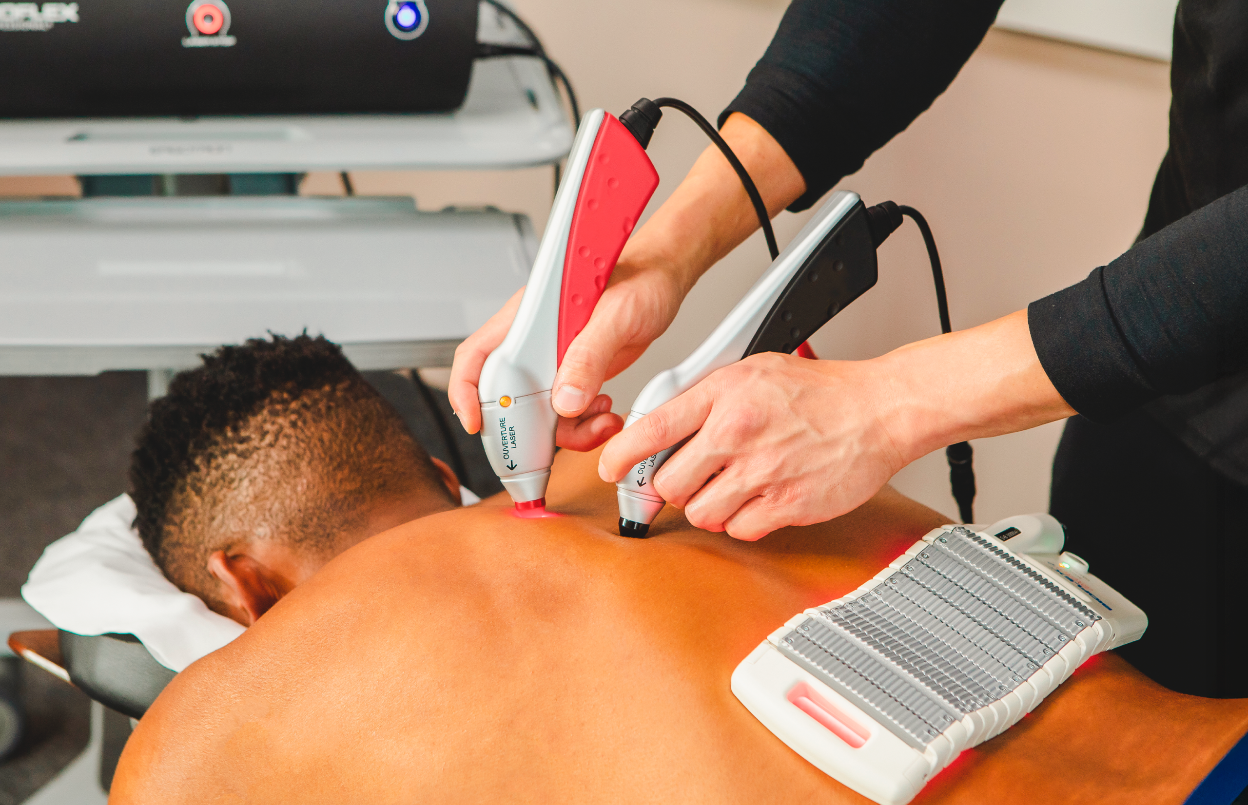
After yellow, the next wavelength in the visible spectrum now falls into what we typically use within our industry, photobiomodulation, red and near infrared. We have used PBMT extensively to treat, or a better word would be hasten, the resolution of ecchymoses. If you look online on using red or near infrared wavelengths for bruises and nine out of ten of the top results would point toward marketing efforts by various PBMT device manufacturers. Truth is, unlike with the shorter wavelengths of light, there probably are no pigments directly involved in ecchymoses progression that PBMT can target. And yet it seems to work. Case in point are two examples below:
Both images show bruises, the upper images are already in advanced stages noting the changing of colour to green then yellow with a few areas that still have a purplish hue. The lower images show a more recent ecchymoses, with all the colours associated with bruises present. For both cases, treatment was initiated using our standard parameters, only 4 treatments were applied daily but the results are noticeable.
As with PBMT, no one knows as of yet the exact mechanism by which the various forms of visible light therapy affect healing, even with the treatments mentioned earlier (blue light phototherapy, PDL and IPL). Cytochrome C-Oxidase, complex 4 in the mitochondrial electron transport chain responsible for transferring an electron from Cytochrome C to Oxygen and eventual production of our energy molecule, Adenosine Triphosphate, and our best friend in PBMT, is a pigmented protein. Copper (CuA) and heme based pigments (Hemea and hemea3) in Cytochrome C-Oxidase are most responsive to the wavelengths we use. CuA has absorption bands at about 480 nm, 530 nm, and even one in the infrared spectrum at about 830 nm.1 Literature has shown that although these absorbances reflect a blue/purple color in the lab, in an intact Cytochrome C oxidase the hemes hide the CuA absorbances with their own absorbances. Hemea has been found to have absorbances at 446 nm and 605 nm. Hemea3 has absorbances at 441 nm, 611 nm, and 656 nm.1 The absorbances for both hemes mean the reflected light gives the hemes a reddish/purple color. These pigments when exposed to the appropriate wavelengths of light mentioned changes their molecular structure and releases Nitric Oxide in return for Oxygen. Note that blue wavelengths (441, 446 and 480 nm) and green (530) are also well absorbed by CCO, why don’t we use them? We could use them of course, but the blue wavelengths cannot penetrate tissues deeply, and green is well absorbed by hemoglobin in red blood cells, which does not have a mitochondrion. So for surface treatments, they are useful, but for deeper tissues, deeper than a few mm, they are wasted.
Another question would be then why does PDL and IPL treatment used green light lasers? PDL and IPL, although still a member of the Photomedicine bandwagon, does a different activity called Photothermolysis, wherein light basically hastens the disintegration of hemoglobin to bilirubin. With hemoglobin most responsive to the green wavelengths, using high intensity light sources like lasers requires rapid pulsed treatments and cooling mechanisms to minimize surrounding tissue damage, and is still dependent on circulation to manage edema and inflammation.
They may not interact directly with the breakdown pigments present in ecchymoses, but PBMT may have a different, but more systemic, effect that aids ecchymoses, and that is the localized improvement in blood flow in the area of treatment. The improved blood flow is due to the unbinding of Nitric Oxide from Cytochrome C Oxidase. Nitric Oxide being a potent vasodilator, improves oxygenation in the area, washes out localized bilirubin pigments into the systemic circulation, and delivers macrophages which will sweep broken down red blood cells and hemosiderin into the lymphatic circulation. Further PBMT encourages collagen production, so any microscopic injury bought about by the injury can also be repaired. Finally, PBMT has an anti-inflammatory effect not directly related to pigment breakdown. So although no one has directly compared the efficacy of PBMT over PDL and IPL, the results should basically be the same.
REFERENCES:
- Hellwig, P., Soulimane, T., Buse, G., and Mantele, W. 1999. Electrochemical, FTIR, and UV/VIS Spectroscopic Properties of the ba3 Oxidase from Thermus thermophilus. Biochemistry, 38: 9648-9658.